Introduction
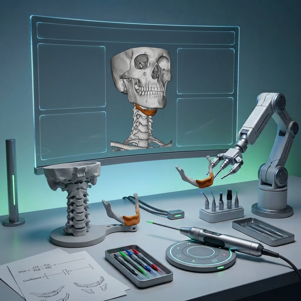
The study aims to develop and test computer-based diagnostic tools for evaluating masseter muscle segmentation on ultrasonography images using deep learning algorithms. The masseter muscle plays a crucial role in masticatory function and its thickness can impact facial morphology and occlusal factors. Ultrasound imaging is beneficial for examining these muscles non-invasively.
Materials and Methods
- Data Collection: The study involved 388 ultrasonographic images of the masseter muscle from anonymous adults. These images were labeled using the polygonal type labeling method with the CranioCatch labeling program.
- Data Set Division: The images were divided into training (312), verification (38), and test (38) sets.
- Deep Learning Model: The artificial intelligence model was developed using the PyTorch U-Net architecture, which is effective in biomedical segmentation.
Results
The U-net model successfully detected and segmented all test images. The success metrics, including F1, sensitivity, and precision, all achieved a perfect score of 1.0.
Discussion
Ultrasound imaging is a reliable diagnostic tool for musculoskeletal examinations. The deep learning approach, specifically using the U-net architecture, showed high precision and recall in segmenting masseter muscles. The study emphasizes the potential of AI in automating quantitative assessments and improving diagnostic efficiency. However, it acknowledges limitations such as the need for larger data sets and further exploration of different architectures.
Conclusion
AI demonstrates significant potential in automating the segmentation of masseter muscles on ultrasonography images, aiding medical practitioners by reducing diagnostic time and effort. Future research should focus on expanding data sets and refining AI models for even better performance.