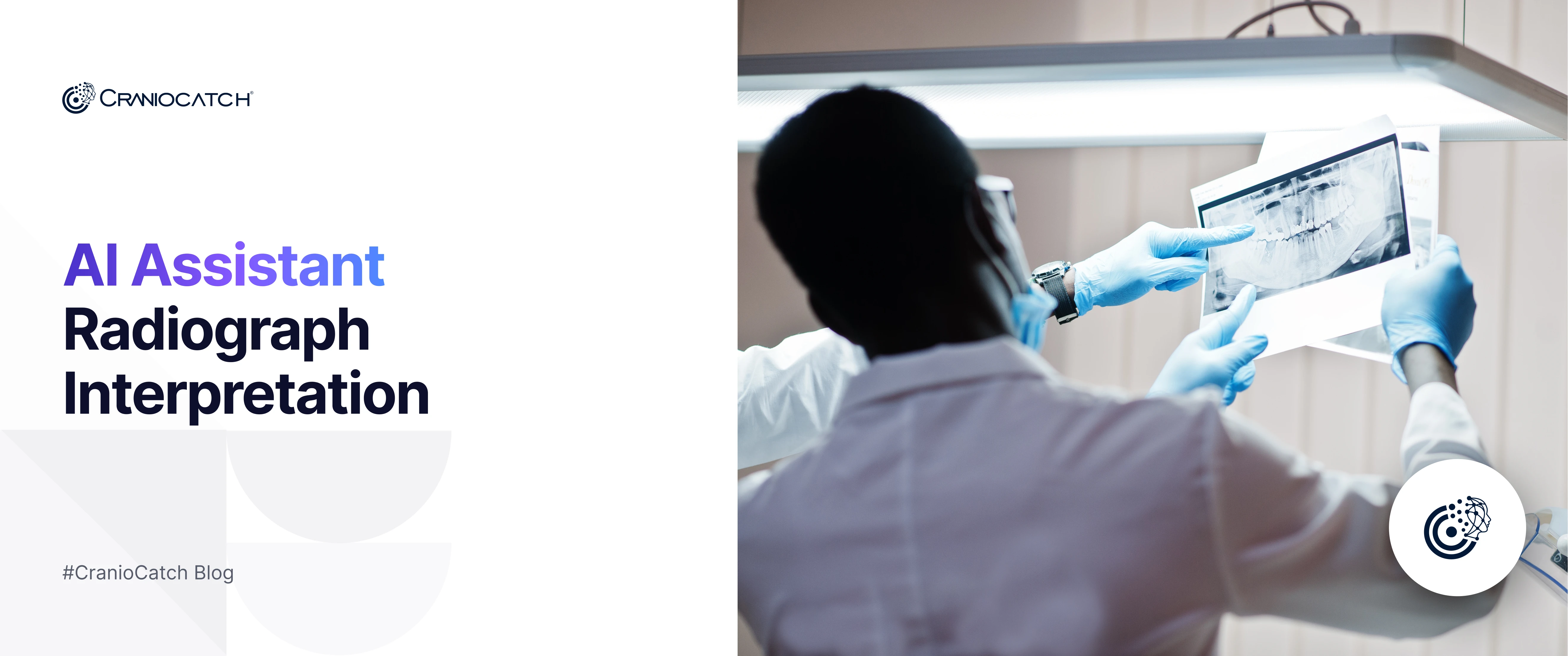
AI-Assisted Dental Radiograph Interpretation: A New Era
AI dental radiography interpretation refers to the use of artificial intelligence and automated image analysis technologies to read and analyze dental X-rays (radiographs) with greater speed and consistency than traditional manual review. These AI dental X-ray readers use sophisticated algorithms — often powered by convolutional neural networks (CNNs) — to detect and highlight structures and potential abnormalities such as caries, bone loss, calculus, periodontal issues, and other pathologies on intraoral or panoramic radiographs.
This automation supports dentists by enhancing diagnostic accuracy, reducing human error, standardizing findings, and enabling faster workflow efficiency while still leaving clinical decision-making in the hands of trained professionals
How AI Interprets Dental X-Rays
AI systems analyze grayscale dental radiographs by applying advanced image processing techniques that transform raw pixel data into meaningful clinical information. First, the algorithm preprocesses the image to enhance contrast and reduce noise, extracting patterns that help distinguish teeth, bone, and soft tissues. Deep learning models then detect and segment key anatomical landmarks — such as individual teeth, root contours, and jaw structures — using convolutional networks trained on large annotated datasets.
AI can also apply color-coded overlays on the original gray-scale radiographs to visually highlight areas of interest, such as suspected caries, restorations, or anatomical features, making subtle anomalies easier to interpret and communicate. This blend of segmentation, landmark detection, and visual augmentation supports faster, more accurate, and more consistent radiographic diagnosis.
Why AI Matters in Modern Dental Diagnostics
AI dramatically improves diagnostic accuracy in dental imaging by detecting subtle signs of caries and other conditions with higher sensitivity and consistency than traditional methods, enhancing clinical confidence and reducing interpretation time.
Its ability to enable early detection of both dental and systemic diseases during routine exams helps initiate timely interventions, improving long-term patient outcomes and enabling personalized care planning.
Types of Dental Radiographic Imaging Used in AI Systems
Dental radiography includes multiple diagnostic imaging dental techniques — from simple X-rays to advanced 3D scans — that AI systems use to analyze and improve clinical interpretation. These imaging types vary in scope, resolution, and purpose, offering a foundation for automated analysis and deep learning-based diagnostics
Periapical Radiographs
Periapical X-rays focus on a small region, capturing detailed images of individual teeth from root to crown. They are essential for detecting tooth decay, root abscesses, and bone loss around specific teeth, and serve as high-resolution input data for AI models trained to identify fine features and abnormalities on radiographs.
Panoramic Radiographs
Panoramic radiographs provide a broad dental imaging view of the entire upper and lower jaws, including all teeth, jaw joints, and supporting bone structures. This wide-field imaging helps identify orthodontic issues, impacted teeth, and larger structural anomalies that AI can analyze for automated detection and comprehensive review.
Cephalometric Analysis
Cephalometric analysis involves side-profile X-rays that capture facial and craniofacial structures, emphasizing anatomical landmarks used in orthodontic diagnosis and treatment planning. AI can recognize and measure these landmarks to assist in assessing jaw relationships and growth patterns.
CT & Computed Tomography in Dentistry
Computed tomography (CT) imaging — especially cone-beam CT (CBCT) — offers detailed three-dimensional views of dentomaxillofacial anatomy, capturing bone density, nerve pathways, and complex structures invisible on 2D X-rays. AI systems analyze these 3D datasets to enhance precision in implant planning, surgical navigation, and complex diagnosis.
AI Technologies Behind Dental Radiography Interpretation
Deep Learning in Dental Imaging
Deep learning forms the core of modern AI dental imaging, using neural networks like CNNs (Convolutional Neural Networks) that learn directly from thousands of radiographs to perform automated image analysis without manual feature engineering. These models classify images, detect regions of interest, and segment teeth and pathologies with high precision by learning visual patterns associated with anomalies and anatomical structures. Deep learning has been successfully applied to panoramic and periapical X-rays, enabling tasks such as tooth detection, identification, and abnormality classification across diverse dental imaging modalities.
Image Processing & Feature Recognition
Image processing techniques enhance and prepare raw radiographic images for analysis by reducing noise, improving contrast, and extracting meaningful features that AI systems can interpret. After preprocessing, AI uses advanced feature recognition such as object detection and segmentation to isolate structures like individual teeth, bone boundaries, and lesions. These recognized features feed into diagnostic workflows, allowing automated systems to highlight caries, periapical lesions, and other dental conditions, making dental radiography interpretation faster and more consistent
Dental Conditions Detected Using AI Radiographic Interpretation
Tooth Decay and Cavities
AI systems have been shown to reliably detect tooth decay / cavities on dental radiographs by analyzing patterns in pixel intensity that correlate with demineralization and lesion presence. Deep learning models trained on large datasets can automatically identify early and interproximal caries, improving sensitivity and diagnostic consistency compared to unaided assessment. These AI tools analyze data from bitewing and panoramic X-rays to highlight areas of decay that might otherwise be missed, supporting timely preventive care and reducing treatment delay.
Periodontal Disease & Bone Loss
AI-based algorithms are highly effective at detecting periodontal disease by quantifying bone loss around teeth on radiographic images. Advanced deep learning ensembles can measure alveolar bone levels, flagging radiographic bone loss with high accuracy and matching expert clinicians in diagnostic performance. Such models assist in determining periodontitis severity and may facilitate earlier intervention before significant structural damage occurs.
Maxillofacial Pathologies
AI systems are increasingly applied to identify maxillofacial cysts and other structural abnormalities on dental scans, including panoramic images. Convolutional neural networks can segment and detect lesions such as dentigerous cysts and potentially guide clinicians toward further evaluation of suspicious bone or soft-tissue pathology. Ongoing research aims to improve lesion-level accuracy for clinical use, expanding the role of AI beyond common dental conditions to broader maxillofacial diagnostic support
Who Uses AI Dental Radiography Interpretation?
Dentists & Dental Clinics
Dentists and dental clinics / dental practices are the primary users of AI dental radiography interpretation tools to support clinical decision-making. These AI systems assist clinicians in identifying dental conditions such as cavities, periodontal issues, and other pathologies on X-rays with improved consistency and speed, helping reduce human interpretation variability across providers in busy practices. AI is increasingly integrated into routine radiographic assessment workflows to enhance diagnostic efficiency and accuracy in clinical settings.
Dental Assistants & Hygienists
Dental assistants and dental hygienists also use AI-assisted radiographic interpretation as part of patient care support, particularly to communicate findings and support dentists’ evaluations. AI helps hygienists explain radiographic results to patients and ensures consistent interpretation among team members, reinforcing collaborative diagnosis and patient education within the dental care team
Benefits and Limitations of AI in Dental Radiology
Benefits for Diagnostic Accuracy
AI significantly enhances diagnostic accuracy in dental radiology by consistently detecting subtle radiographic patterns associated with caries, periodontal changes, and structural abnormalities. Deep learning models improve early detection by identifying lesions at initial stages that may be overlooked during manual interpretation, reducing false negatives and supporting more objective, reproducible diagnoses across clinicians.
Impact on Patient Outcomes
Improved diagnostic precision directly influences patient outcomes, as earlier and more accurate identification of dental conditions enables timely intervention, minimally invasive treatment, and better long-term oral health management. However, AI remains a clinical decision-support tool rather than a replacement for professional judgment, requiring expert oversight to address data bias, imaging variability, and ethical responsibility in patient care.
The Future of AI in Dental Radiography Interpretation
The future of AI-driven dental imaging points toward increasingly automated diagnostics that will deepen integration with clinical workflows and expand into maxillofacial radiology beyond conventional dental X-rays. Research shows that AI systems are already matching or exceeding human performance in dental findings detection across diverse datasets, processing images far faster while maintaining high sensitivity and specificity — a trend that will continue as models and clinical validation improve.
Emerging developments will likely combine automated diagnostics with advanced 3D imaging like CBCT, enabling more precise interpretation of complex anatomical structures and pathologies within the dentomaxillofacial region. AI is also poised to enhance clinical decision support by integrating radiographic data with patient records and predictive analytics, helping tailor individualized treatment plans and improve care outcomes.
As AI tools evolve, the role of dental specialists — including radiologists — will shift toward overseeing and validating AI outputs, ensuring ethical use and clinical relevance while leveraging these technologies to streamline diagnostic workflows, reduce variability, and support earlier intervention


 Contact Us