Dental Imaging Essentials: A Comprehensive Guide for Radiologists

- What is Dental Imaging?
- Role of Dental Imaging in Dentistry
- Types of Dental Imaging Techniques
- How are Dental X-Rays Used for Diagnosis?
- Role of Cone Beam CT in Dental Radiography
- How Do Digital Dental Imaging Technologies Improve Patient Care?
- Cariology and Caries Management
- Digital Technology in Endodontic Practice
- FAQ
Dental imaging plays a crucial role in modern dentistry, providing valuable insights into the oral health of patients. By utilizing various imaging techniques, dentists can accurately diagnose dental conditions and create effective treatment plans tailored to individual needs.
What is dental imaging?
Dental imaging involves capturing images of the teeth and surrounding structures to diagnose and monitor oral health. This can include bitewing x rays to detect cavities between teeth, full mouth x rays to assess overall dental health, and healthy teeth x rays to track changes over time.
Bitewing x rays are commonly used in dentistry to provide a detailed view of the upper and lower teeth. By taking these images, dentists can identify issues such as decay, gum disease, and bone loss that may not be visible during a regular exam. Xray teeth help dentists provide accurate diagnoses and develop treatment plans to maintain oral health.
Definition and importance of dental imaging
Dental imaging refers to the process of capturing detailed images of teeth, bones, and soft tissues in the mouth using specialized equipment. It is an essential tool in modern dentistry, allowing dentists to diagnose dental conditions and monitor oral health over time. Techniques like dental x-rays, 3D dental imaging, and dental radiographs provide dentists with critical information that is not visible during a regular exam. With advancements in dental imaging technologies, dentists can now utilize dental digital imaging to ensure more precise and accurate diagnoses, leading to better treatment outcomes. In addition, dental imaging software plays a key role in enhancing the clarity and utility of these images, making diagnosis faster and more reliable.
Role of dental imaging in dentistry
Dental imaging plays a key role in various aspects of dentistry, including diagnosis, treatment planning, and monitoring of oral health. It enables dentists to identify problems such as tooth decay, bone loss, and impacted teeth, leading to more effective dental care.
Benefits of utilizing dental imaging
The benefits of dental imaging are manifold. It helps in detecting dental diseases at an early stage, reducing the risk of complications. Additionally, dental imaging aids in determining the appropriate treatment approach, resulting in improved patient outcomes and overall oral health.
Types of dental imaging techniques
In modern dentistry, there are several dental imaging techniques used to visualize oral structures. Intraoral imaging, such as bitewing and periapical dental x-rays, provides detailed images of the teeth and supporting bone structure, ideal for diagnosing issues like decay and periodontal disease. On the other hand, extraoral imaging techniques, like panoramic dental radiography and cone beam CT, offer a comprehensive view of the entire jaw and facial bones. Each of these techniques utilizes different dental imaging technologies, tailored to the specific diagnostic needs of the patient. Furthermore, advanced 3D dental imaging techniques are used in more complex cases, such as implant placement and orthodontic treatment, ensuring greater accuracy in treatment planning.
Intraoral imaging methods
Intraoral imaging methods involve capturing detailed images of the teeth and supporting structures within the mouth. This includes techniques such as periapical x-rays, bitewing x-rays, and occlusal x-rays, which provide valuable information about individual teeth and specific dental structures.
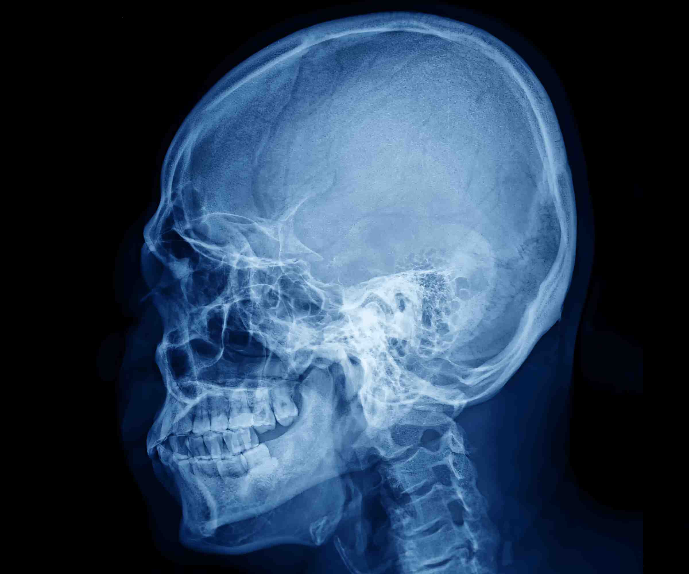
Extraoral imaging techniques
Extraoral imaging techniques are used to capture broader images of the entire oral and maxillofacial region. This includes panoramic x-rays and cone beam computed tomography (CBCT), which offer a more comprehensive view of the dental structures, facilitating accurate diagnosis and treatment planning.
Comparison between different types of dental imaging
Each type of dental imaging technique has its own advantages and limitations. While intraoral imaging methods are ideal for detailed views of individual teeth, extraoral techniques provide a broader perspective of the entire oral cavity, aiding in the evaluation of overall oral health.
How are dental x-rays used for diagnosis?
Dental x-rays are critical tools in the diagnosis and treatment planning of various dental conditions. By using dental radiographs, dentists can detect cavities, bone loss, impacted teeth, and other abnormalities that are not visible during a routine exam. Dental x-ray machines capture high-quality images that allow for early detection of dental issues, reducing the risk of complications. With the help of dental digital imaging and dental imaging software, these x-rays are quickly processed and analyzed, ensuring a more efficient diagnostic process. Radiology in dentistry plays a vital role in identifying issues that require immediate intervention, ultimately improving patient care.
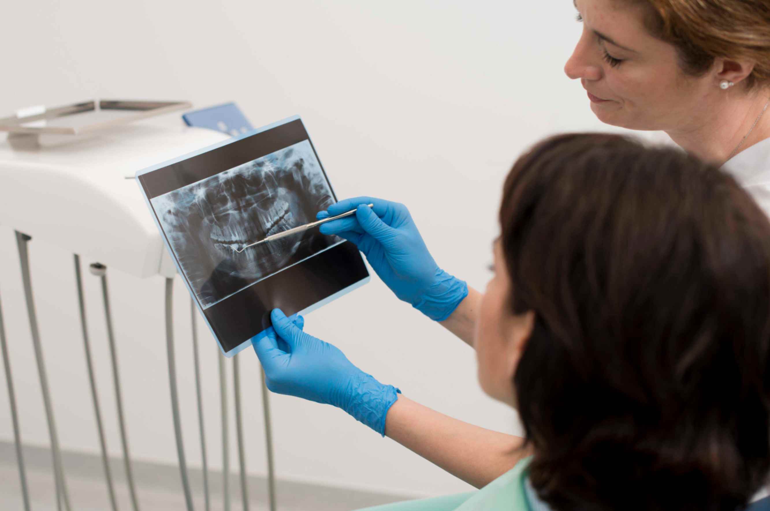
Process of taking and analyzing dental x-rays
Dental x-rays are commonly used for diagnosing dental conditions by capturing images of the teeth and surrounding structures. The process involves positioning the x-ray machine to take specific images, which are then analyzed by dentists to identify any abnormalities or signs of dental disease.
Common dental conditions detected by x-rays
Dental x-rays are effective in detecting various dental conditions, including tooth decay, bone loss, and dental infections. They also help in identifying impacted teeth, assessing the extent of dental decay, and monitoring the progress of ongoing dental treatments.
Advancements in digital dental radiography
Advancements in digital dental radiography have revolutionized the field of dental imaging by providing high-quality images with reduced radiation exposure. Digital x-rays offer enhanced clarity and detail, allowing for more precise diagnosis and improved patient care.
Role of cone beam CT in dental radiography
Cone beam CT (CBCT) has revolutionized dental radiography by providing 3D images of the teeth, jaw, and surrounding structures. This technology allows dentists to visualize complex cases in greater detail, offering better precision for procedures like dental implants and orthodontics. Unlike traditional dental x-rays, CBCT scans capture a full 3D image, making it easier to assess bone density, root canal morphology, and the exact position of impacted teeth. As an advanced form of dental imaging, CBCT is increasingly used in surgical planning, allowing for safer and more predictable outcomes in dental treatments.
Understanding cone beam computed tomography (CBCT)
Cone beam CT is a specialized imaging technique that generates 3D images of the dental structures, offering a comprehensive view of the teeth, jaw, and surrounding tissues. It provides detailed information for complex dental procedures and precise treatment planning.
Applications of cone beam CT in dentistry
Cone beam CT is widely used in various dental specialties, including oral surgery, orthodontics, and implant dentistry. It helps in assessing bone quality, identifying the exact position of dental implants, and analyzing the relationship between teeth and surrounding structures.
Benefits and limitations of cone beam CT
The benefits of cone beam CT include improved accuracy in diagnosis, reduced radiation exposure compared to traditional CT scans, and enhanced treatment outcomes. However, its limitations lie in cost considerations and the need for specialized training to interpret the complex 3D images accurately.
How do digital dental imaging technologies improve patient care?
Digital dental imaging has significantly enhanced the quality of care provided to patients. Unlike traditional dental radiography, which relied on film, dental digital imaging produces clearer, more detailed images with less radiation exposure. This advancement in dental imaging technologies allows for quicker image acquisition and analysis, leading to faster diagnoses and more efficient treatment planning. With the integration of dental imaging software, dentists can magnify and manipulate images to focus on specific areas, ensuring more accurate treatment decisions. Ultimately, these innovations in imaging dental solutions contribute to improved patient outcomes by enhancing the precision and speed of dental care.
Advantages of digital x-rays over traditional film-based radiography
Digital x-rays offer numerous advantages over traditional film-based radiography, including faster image acquisition, easy storage and retrieval of images, and the ability to enhance images for better visualization. They also reduce radiation exposure for patients and dental staff.
Enhanced accuracy and efficiency through digital dental imaging
Digital dental imaging technologies improve the accuracy and efficiency of diagnosis and treatment planning by providing clear, high-resolution images. Dentists can easily magnify and manipulate digital images to focus on specific areas of interest, leading to more precise dental care.
Integration of digital dental imaging in modern dentistry
With the increasing digitization of healthcare, digital dental imaging has become an integral part of modern dentistry. It allows for seamless communication between dental professionals, facilitates interdisciplinary collaborations, and enhances the overall quality of patient care.
Cariology and Caries Management
When it comes to managing caries, dental x rays play a crucial role in diagnosis and treatment planning. Intraoral radiography is commonly used to detect teeth cavities and assess the health of the surrounding bone. Different types of dental x ray machines can be utilized, including bitewings x ray and periapical x rays, to capture detailed dental x ray images of the upper and lower teeth. By using x-rays to evaluate the supporting bone and developing teeth, dentists can provide a comprehensive diagnostic imaging approach to dental care.
Bitewing x rays show the bitewings xray of the mouth, while full mouth series x rays provide a complete view of the front teeth and occlusal x ray of the jaw. These x-rays help in identifying healthy teeth, as well as fractures or abnormalities in the teeth x ray machine. With advancements in dental radiology and new technology, x-rays taken in a dental office now expose patients to less radiation while providing detailed information about their medical and dental history.
Optical Coherence Tomography
Optical Coherence Tomography (OCT) is a non-invasive imaging technique that provides high-resolution cross-sectional images of the internal structures of biological tissues. While traditional dental x-rays such as bitewing x rays and full mouth series of x rays are still commonly used for diagnostics in dentistry, OCT offers a more detailed view of the teeth and jaw bone. This type of x-ray helps dentists detect issues such as teeth cavity or periapical xray – from the crown to the root – without the need for radiation exposure concerns.
Unlike cephalometric radiograph or periapical xray dental, which are types of extraoral x-rays, OCT is considered an intraoral x-ray that focuses on capturing images of your mouth at a higher resolution. This type of x-ray helps in planning dental work, such as restorative procedures, by providing x-ray image of the gland and head and neck region. With OCT, dentists can get a clearer view of the lower teeth in one area or front teeth xray for better diagnostics.
Digital Technology in Endodontic Practice
Digital technology in endodontic practice has revolutionized the way dentists diagnose and treat patients. With the use of x-ray machines, such as bite wing x-rays and full mouth x-rays, dentists can get a clear picture of the status of developing teeth and detect any cavities or issues with the jaw bone. These dental radiographs are digitally stored, allowing for easy access and comparison during follow-up appointments. This new technology is essential for making a precise diagnosis and ensuring the overall oral health of the patient.
One of the most common types of dental x-rays is the bitewing x-ray, which shows details of the upper and lower teeth in relation to each other. Another important type is the periapical x-ray, which focuses on a specific tooth from its crown to the root. By using this new technology, dentists can check for any abnormalities or concerns inside the mouth and address them promptly.
Graphics and Imaging
Dental x-rays help dentists get a closer look at the status of developing teeth and identify potential problems such as teeth cavities. There are two main types of intraoral x-rays: full mouth x-rays and bitewing x-rays. It is recommended to get x-rays every six months to check the health of your teeth. A periapical x-ray shows a detailed image of your teeth and jaw, while a full mouth x-ray accounts for approximately 18 images of your mouth while blurring around the head.
Conventional dental x-rays detect issues such as teeth cavities and jaw bone abnormalities. A bitewing x-ray specifically focuses on the front teeth and normal teeth. These x-rays are essential for making a diagnosis and identifying any concerns early on. If you are concerned about radiation exposure, talk to your dentist about ways to minimize it during x-ray procedures. Remember, regular x-rays can help maintain healthy teeth and gums.
FAQ
What are the three types of dental images?
There are three types of dental images that dentists commonly use to assess the health of a patient's teeth and jaws. One type is the bitewing x-ray, which is used to check the status of developing teeth cavities. Another type is the full mouth x-ray, which provides a comprehensive view of all the teeth in the mouth. And the third type is the x-ray of front teeth, which focuses on the front teeth and jaw bone.
How does dental imaging work?
How does dental imaging work? Dental imaging involves various types of x-rays to capture images of different parts of the mouth. Bitewing x-rays focus on the teeth cavities and can detect normal teeth. A full mouth x-ray provides a comprehensive view of all the teeth, while a front teeth x-ray focuses on the front area. The x-ray of the jaw bone helps in diagnosing issues with the jaw. The radiation beam emitted by the pa x-ray dental machine captures detailed images of healthy teeth.
What is the latest dental imaging?
For the latest dental imaging, dental x-ray bitewings and full mouth x-rays are commonly used to detect teeth cavities and assess the health of front teeth. In addition, x-ray jaw bones and normal teeth x-rays are crucial for identifying any underlying issues. Bitewing x-rays provide detailed images of the teeth and jaw to aid in diagnosis and treatment planning.
What are the disadvantages of dental imaging?
One major disadvantage of dental x-ray bitewing is the exposure to radiation, which can pose risks in the long term. In addition, full mouth x-rays can be time-consuming and costly for patients. Another drawback is the difficulty in detecting teeth cavities in areas not covered by bitewing x-rays.
Furthermore, jaw x-rays may not always provide a clear view of the entire jaw bone, leading to potential inaccuracies in diagnosis. Additionally, while x-rays of front teeth can be useful for detecting issues in that area, they may not capture potential problems in other parts of the mouth. Overall, there are limitations to the scope of dental imaging techniques.
Why is dental imaging important?
Having dental imaging done regularly is crucial for maintaining good oral health. Bitewing x-rays can detect teeth cavities early, while full mouth x-rays provide a comprehensive view of the entire mouth. A jaw x-ray can reveal issues with the jaw bone, and front teeth x-rays can identify abnormalities in the front teeth.
What is dental 3D imaging?
Dental 3D imaging is a technique that allows dentists to capture detailed images of a patient's teeth, jaw, and surrounding structures in three dimensions. Unlike traditional x rays, which only provide a two-dimensional view, 3D imaging offers a more comprehensive look at a patient's oral health. This technology can be especially useful for detecting issues such as teeth cavities, abnormalities in the jaw bone, and full mouth x-rays.
One common type of x-ray used in dental imaging is the bitewing x-ray, which focuses on the teeth in the upper and lower jaws. Bitewing x-rays are helpful for examining the front teeth and detecting pa x-ray dental decay between adjacent teeth. These x-rays can provide a close-up view of healthy teeth and help dentists identify potential issues early on.
What is a digital imaging in dentistry?
What is a digital imaging in dentistry? Digital imaging in dentistry refers to the use of technology like x-ray teeth to capture detailed images of a patient's oral health. This includes full mouth xray, bitewing x ray, front teeth x ray, and jaw x ray to assess conditions such as teeth cavity x ray and normal teeth xray.
One common type of x-ray used in dentistry is the dental x ray bitewing, which gives a detailed view of a specific area of the mouth, typically the molars and premolars. This helps dentists detect issues like cavities and decay. Another important x-ray is the pa x ray dental, which provides a comprehensive view of the entire mouth, including the x ray of front teeth and x ray jaw bone.


 Contact Us