What is Digital Imaging Dental? Benefits and Applications in Modern Dentistry

Digital imaging dental is a transformative technology in the field of modern dentistry, enabling precise, efficient, and patient-friendly diagnostics. By replacing traditional film-based methods, digital dental imaging enhances accuracy and reduces exposure to radiation. The applications of this technology span diagnostic, restorative, and surgical treatments, contributing to improved patient outcomes.
This article delves into the essentials of digital imaging dental, exploring its types, benefits, and integration with advanced systems like AI-powered solutions.
What is Digital Dental Imaging?
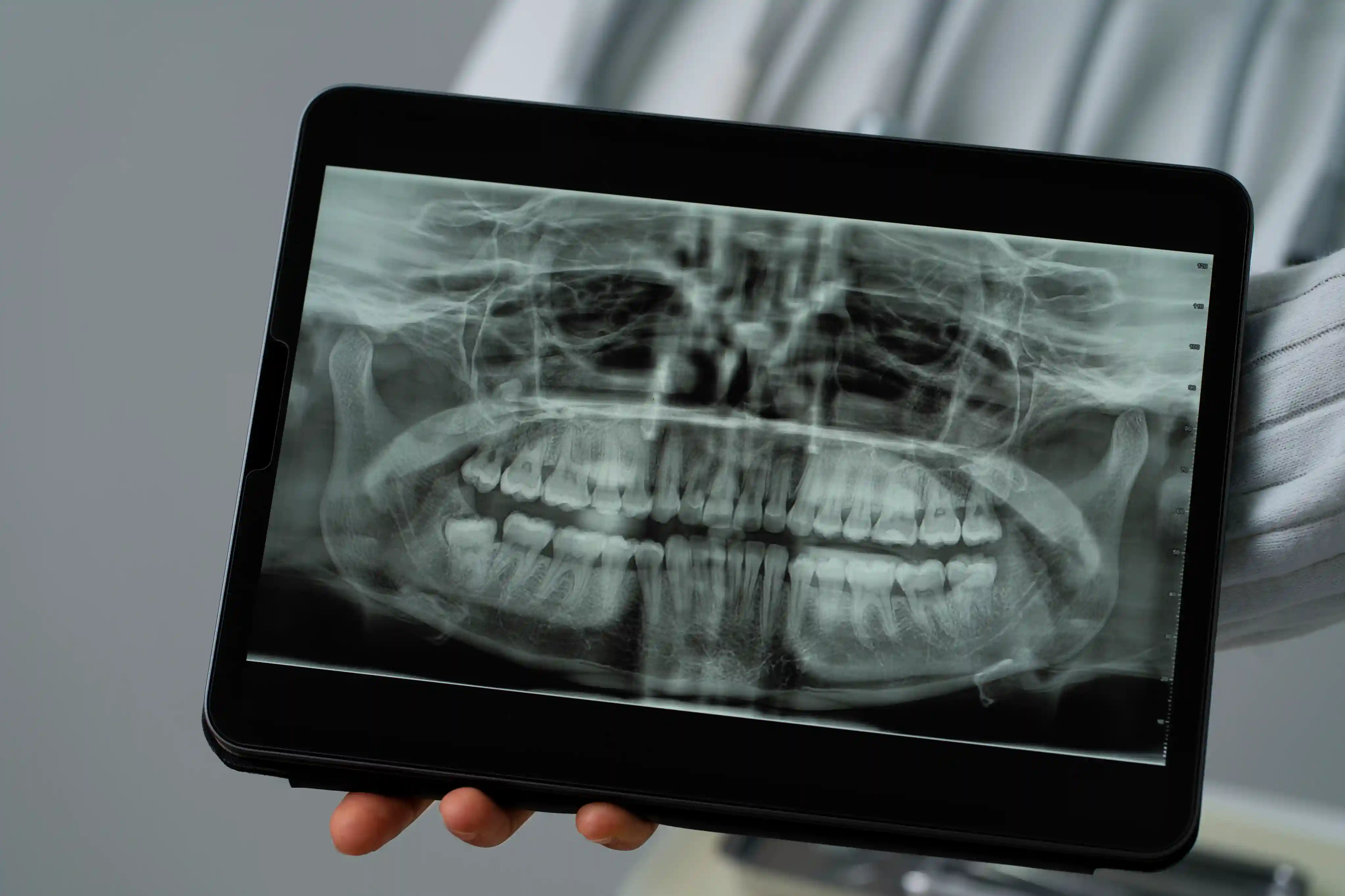
Digital dental imaging refers to the use of digital technology to capture, process, and store dental radiographs. Unlike traditional X-rays, this technique employs sensors and software to produce instant, high-quality images that can be easily shared and analyzed.
The process begins with the placement of a sensor in the patient’s mouth. This sensor captures images, which are then transferred to a computer for immediate review. This method reduces patient radiation exposure while providing superior image clarity, aiding in accurate diagnoses.
Dentists use digital dental imaging systems to identify issues such as cavities, periodontal disease, and jawbone abnormalities. The real-time imaging capability supports better treatment planning, ensuring timely interventions for dental health problems.
How Does a Digital Dental Imaging System Work?
A dental digital imaging system operates by replacing traditional photographic film with advanced electronic sensors. These sensors are sensitive to X-rays and convert them into digital signals, which are processed by specialized software.
1. Image Capture: When X-rays pass through the teeth and gums, the sensor detects variations in density and records the data.
2. Image Processing: The recorded signals are processed into detailed, high-resolution images.
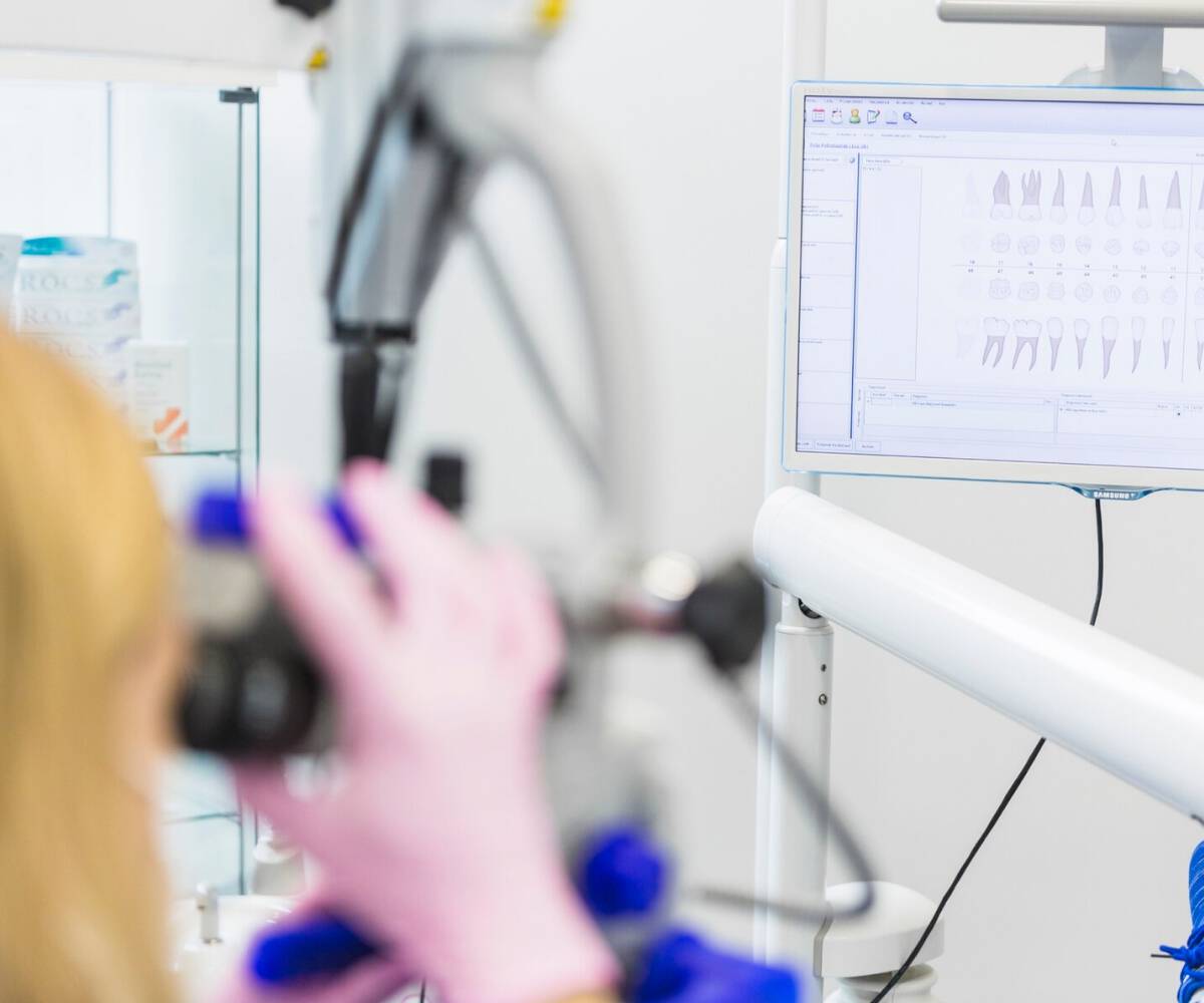
3. Image Storage and Sharing: The digital images are stored electronically and can be easily shared with specialists or included in patient records.
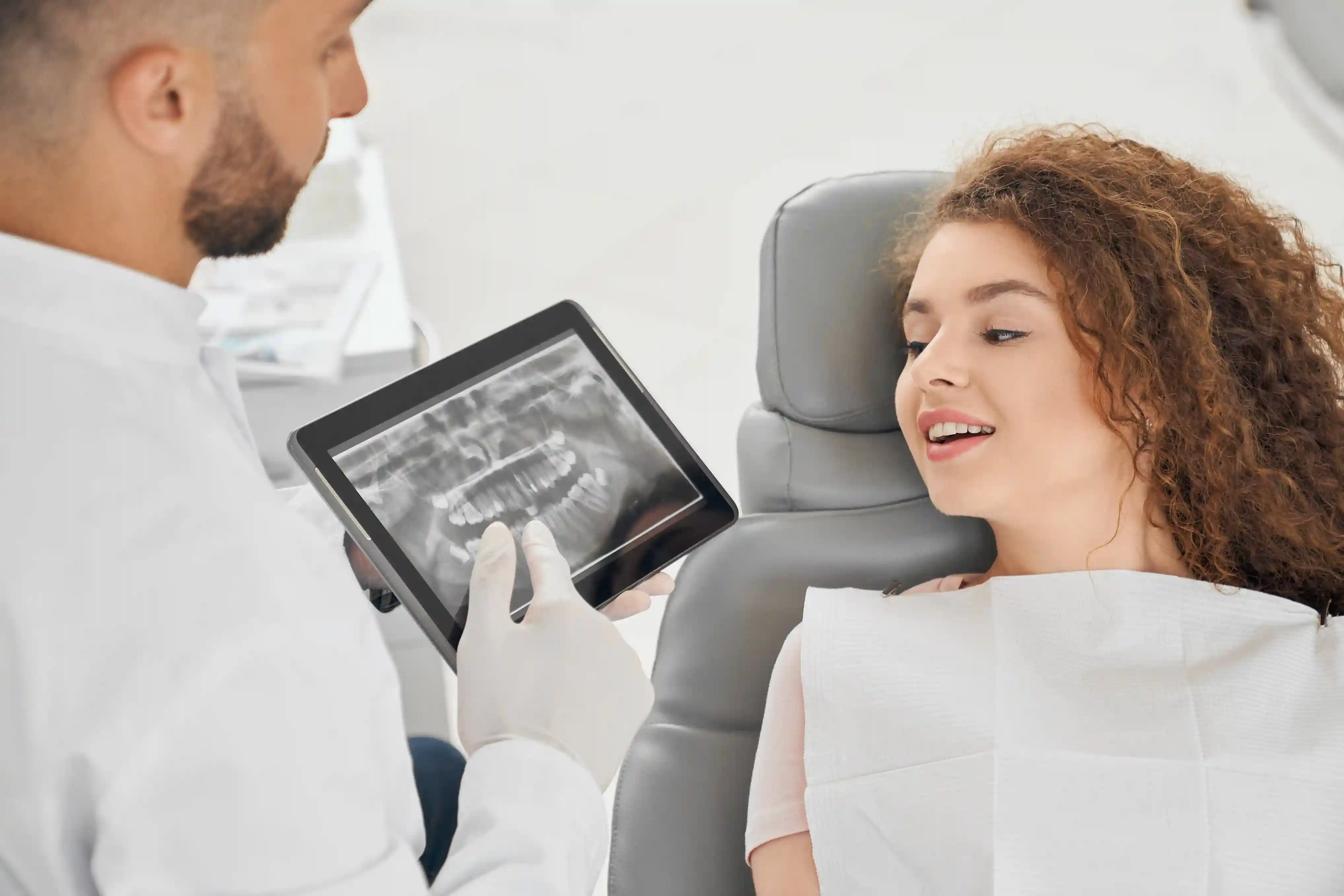
The integration of direct digital imaging dental reduces the time spent waiting for results and enhances collaboration among dental professionals. Furthermore, the system’s compatibility with AI tools supports predictive analytics and personalized treatment recommendations.
Types of Dental Digital Imaging Systems
There are several types of dental imaging systems designed to meet the varying needs of dental practices. Each offers unique features to suit specific diagnostic and procedural applications.
1. Intraoral Digital Imaging Systems: These capture detailed images of individual teeth and surrounding structures. They are commonly used for detecting cavities and assessing root canals.
2. Extraoral Digital Imaging Systems: These provide a broader view of the jaw and skull, making them ideal for orthodontic and surgical planning.
3. 3D Imaging Systems: Technologies like cone beam CT scans offer a three-dimensional view of the oral cavity. For more details, read about cone beam CT scan.
The versatility of dental imaging technologies ensures precise diagnostics, facilitating comprehensive dental care.
Benefits of Digital Dental Radiography
The shift to digital dental radiography brings numerous advantages for both patients and practitioners. Here are some key benefits:
1. Reduced Radiation Exposure: Patients experience significantly less radiation compared to traditional X-rays.
2. Instant Results: Digital images are available immediately, eliminating the need for film development.
3. Enhanced Diagnostics: High-resolution images improve the accuracy of diagnoses and treatment planning.
4. Environmentally Friendly: Eliminates the need for chemical processing, reducing environmental impact.
By leveraging digital radiography in dentistry, professionals can streamline workflows, improve patient satisfaction, and ensure long-term health outcomes.
What are the Applications of Digital Radiographs in Dentistry?
Digital radiographs play a pivotal role in various dental specialties. Their applications include:
- Preventive Care: Identifying cavities, plaque buildup, and early signs of gum disease.
- Orthodontics: Planning braces or aligners with precise measurements.
- Surgical Procedures: Mapping out surgeries such as implant placement or wisdom tooth extraction.
- Prosthodontics: Designing crowns, bridges, and dentures with accurate imaging.
The adaptability of dental digital radiography systems ensures their relevance in diverse dental treatments, contributing to enhanced care and patient outcomes.
Disadvantages of Digital Dental Radiographs
While digital dental radiographs offer numerous benefits, they also have some limitations:
1. Initial Costs: Setting up dental digital imaging systems can be expensive, requiring significant investment in equipment and training.
2. Technical Challenges: System malfunctions or software issues may disrupt workflows.
3. Learning Curve: Dentists and staff may face challenges in adapting to new systems and technology.
Despite these challenges, the advantages of digital dental x ray systems often outweigh the drawbacks, making them a worthy investment for modern dental practices.
AI-Powered Solutions for Dental Digital Imaging Systems with CranioCatch
AI is revolutionizing the field of dental digital imaging systems, and CranioCatch is at the forefront of this innovation. By combining artificial intelligence with advanced imaging, CranioCatch enhances diagnostic precision and treatment outcomes.
1. Automated Diagnostics: AI algorithms analyze images for early detection of dental issues.
2. Improved Workflow: AI reduces the time required for image analysis, allowing practitioners to focus on patient care.
3. Personalized Treatment Plans: AI tools recommend tailored treatment strategies based on patient data.
You can visit our website and contact us to learn more about how AI in dental imaging transforms dentistry.


 Contact Us